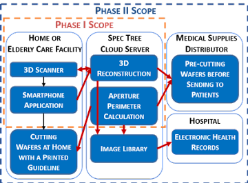
We want to use the Stoma-Wafer Inspection and Fit Tool (SWIFT) to enable higher quality stoma care at a lower cost for people with inestinal or urinary diversion. Better post-operative wafer fitting has been identified as a key area for stoma care improvement. Wafer fitting is typically conducted by trial and error, requiring the re-sizing of the wafer opening due to changes in patient weight, size of the stoma, and shape or condition of the peristomal area. These trials may cause great damages or discomfort for patients and we wish to minimize them.
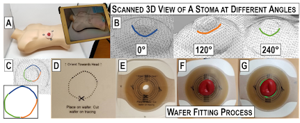
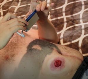
See the image on the corresponding image on the right for photographs and screenshots of our prototype 3D scanning and reconstruction process for a model stoma. Starting with (A), a 3D scan is taken. The system identifies the edges of the stoma to determine wafer cut perimeter. A stoma tracing calculation algorithm is then applied (B, C) once the stoma tracing pro-cess is complete. The results are printed out on a transparent sheet for easy manual cutting (D, E), but it could be done automatically by laser cutters in the future. The guided cut has optimal shape (F), while (G) shows a manual cut made by a current stoma patient. The deviation from the ideal cut in (F) are highlighted in green.
We want to create an application that utilizes the SWIFT concept. Specifically, we wish to be able to take pictures of stomae and accurately calculate the peristomal area for analysis and construction of prototype wafers.